Dbios Anatomical Models are available for Medical , Dental, Homeopathic and Ayurvedic Colleges through out the world. These Models are made of unbreakable fiber glass or plastic which long lasts in your labs. The vivid knowledge of Embryology, Histology or Anatomy models for upcoming Doctors are one of best available in India. For complete list visit download section. We are manufacturers of all kind of Educational Models and charts, from School, Medical Colleges to Technical Institutes. For complete Embryo charts-models download section
RARE Dbios Anatomy Models
AM1 Model of Man or Woman Life Size : 160CM Showing superficial dissection on one side. And other side intact.
AM2 Human Torso with Head life size torso
AM3 Human Torso with Head life size Sexless Height 90 cms.
AM4 Head & Neck (Median Section) : Made of fiber glass, mounted on base.Size : 10″ x 12.3″ / 4″ x 2″
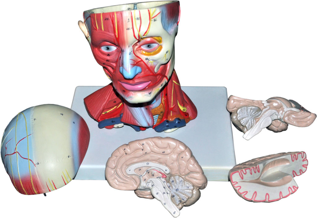
AM5 Head and Neck dissectable:in two parts in L.S. showing superficial dissection on one side and other intact. Half brain can be taken out.
AM6 Brain with Skull
AM7 Human Brain in 4 Parts
AM8 Brain in 8 parts
AM9 Midsagittal Section through the brain extra large showing all details
AM10 Structure of Cerebellum :A Superior View,An Interior View,A Sagittal View
AM11 Sagittal Section of the Medulla Oblongata and Pons Showing the Cranial Nerve Nuclei of Gray Matter.
AM12 The Autonomic Nervous System: showing relationship of different organs to the spinal cord.
AM13 Human Nervous System half the natural size schematic presentation of the central and peripheral system.
AM14 Visual Central Nervous System Pathways.
AM14 (a) Neuron Model Imported.
AM15 Spinal Cork with Spinal Nerves.
AM16 Human Eye in Socket V.S. dissectable in 7 parts.
AM19 Muscular Eye with Orbit – Enlarged about 4-times, Size: 10″x13″x9.1/2″
AM20 Ear Large Size showing External, Middle and Inner Ear Dissectable in four parts.
AM21 Ear Sagittal Section extra large and detailed model
AM22 Ear six time enlarged made from Venyl Rubber.
AM24 Teeth with tongue.
AM23 Tonsils : Pharyngeal, Palatine & Lingual Tonsils.
AM25 Upper and Lower Jaw
AM26 Pituitary Gland Hypothalamus.
AM27 Thyroid & Parathyroid Gland.
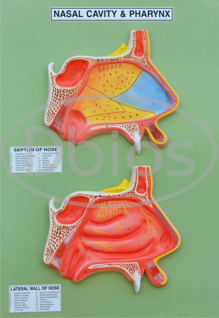
AM28 Nascal Cavity & Pharynx Sagittal section viewed from medial side.
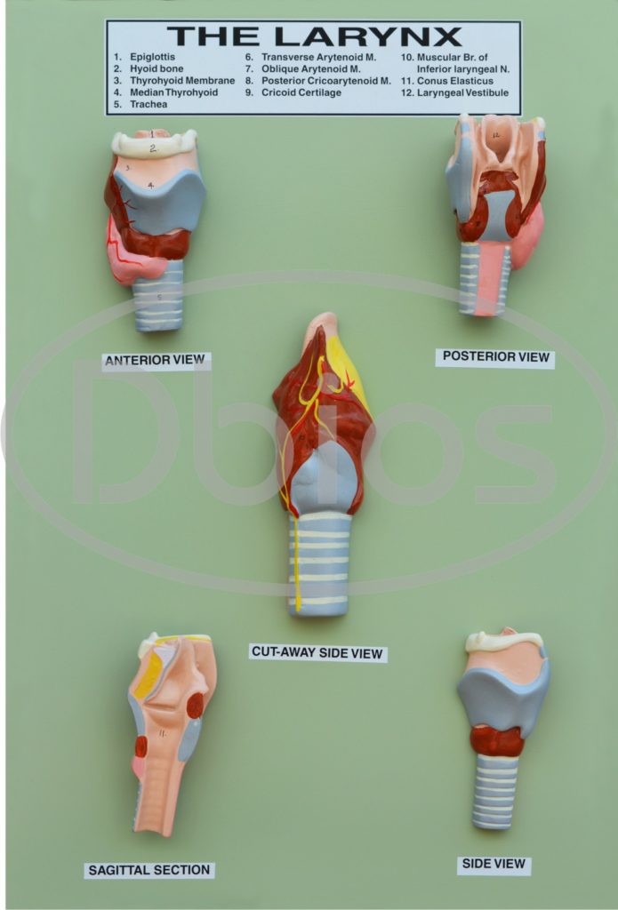
AM29 Larynx Anterior View, Posterior View, Slide View cut away side view and Sagittal Section.
AM30 Larynx Deep side view.
AM32 Pharynx & Larynx Sagittal Section.
AM34 Lungs section with respiratory tract, Bronchial tubes, Arteries & Veins.
AM35 Human Lungs with Heart ;approximately 20″x18″
AM36 The Respiratory System.
AM37 Liver enlarged showing Gall Bladder.
AM38 Liver with Gall Bladder & Pancreas.
AM39 Liver : showing blood supply.
AM40 Pancreas enlarged
AM41 Stomach enlarged with duodenum section
AM42 Spleen Normal size with details.
AM43 Gall Bladder, Pancreas & Duodenum.
AM44 Intestine showing blood supply.
AM45 Intestine Large and Small.
AM46 Rectum (Anal Canal).
AM47 Duct System.
AM48 The Digestive System.
AM49 Heart Enlarged seperable in 4 parts.
AM49 (b)Imported Giant Heart on Diaphragm
AM50 Circulatory System.
AM51 The Hepatic Portal System.
AM52 Schematic Circulatory System.
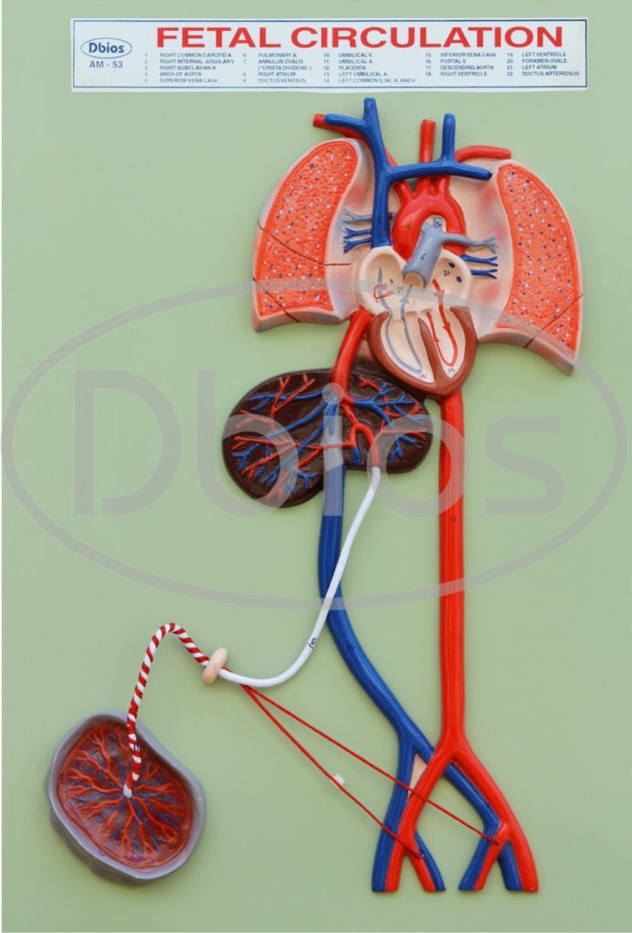
AM53 Fetal Circulation.
AM54 Arteries of the Neck & Arteries of the Neck & Head,
AM55 An Anterior View of the Major Arteries of the Upper Extermity.
AM56 Arteries of the Pelvic Region.
AM57 Arteries of the Right Lower Extremity (Anteri &P)
AM58 Urinary System with Kidneys and Urinary Bladder.
AM59 Kidney enlarged showing Nephrone & glomerulus on board.
AM60 Urinary Bladder Sectio
AM61 Testis Cross Section.
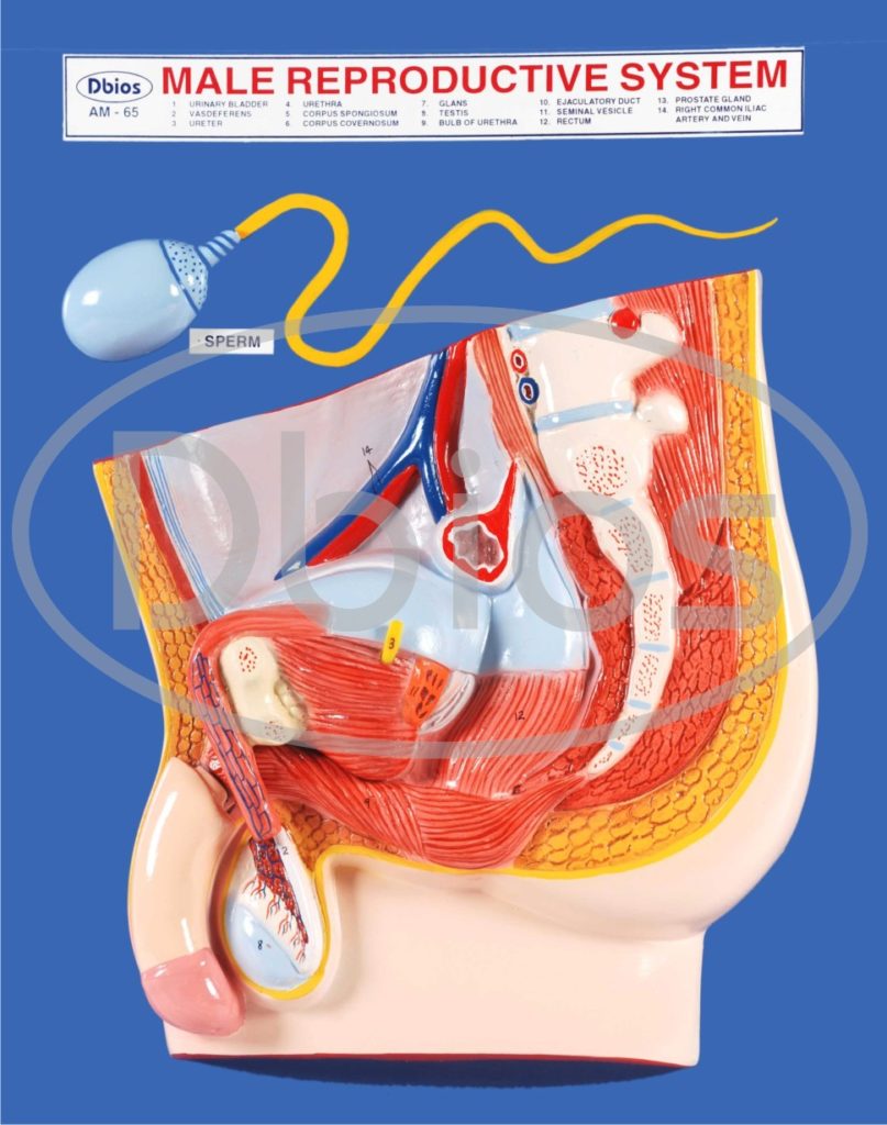
AM62 Penis Cross Section Anterior view
AM63 Structure of the Penis Showing the attachment, blood and Nerve supply.
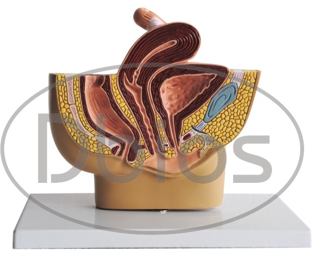
AM64 Female Urethra L.S.
AM65 Skin Cross Section Model 100 times enlarged
RARE HISTOLOGY /MICRO- ANANTOMY MODELS.
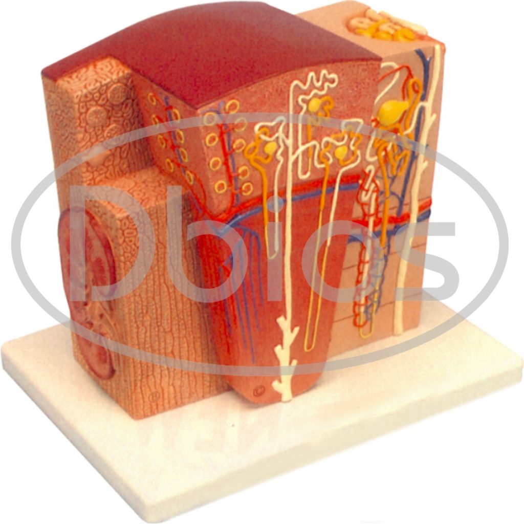
IMP284 MICRO anatomy Kidney
This extremely detailed model shows the morphologic/functional units of the kidney greatly magnified. Six model zones illustrate the following fine-tissue structure that serve the production or urine :
- Longitudinal section of a kidney
- Section of renal cortex and renal medulla
- Wedge-shaped section of a kidney lobe with a diagrammatic depictionof three nephrons with Henle’s loop and didactic/ diagrammatic illustration of the vascular supply
- Diagrammatic illustration of a nephron with a short Henle’s loop and didactic/diagrammatic illustration of the vascular supply
- Diagrammatic illustration of an opened renal corpuscle with nephron and light-microscopic tranverse sections of the proximal, attenuated and distal segments of a renal tubule
- Diagrammatic / didactic illustration of an opened renal corpuscle
- Mounted on a base. 5×25.5×19 cm ; 1.3 kg
IMP 289 Micro Anatomy Muscle Fibre
- The model illustrates a section of a skeleton muscle fibre and its neuromuscular
- end plate magnified approx. 10.000 times. The muscle fibre is the basic element of the diagonally striped skeletal muscle.
- 5x26x18.5 cm.;1.1kg.
IMP286 MICRO anatomy Digestive System
- The model illustrations the structure of the fine tissues of four characteristic
- sections of the digestive system:
- l Oesophagus l Stomach l Small intesting l Large intestine
- The front of the model, from top to bottom, shows a magnified view in
- histological section of the individual sections of the digestive system and their
- fine tissue structures. On the back of the model, highly magnified views of
- didactically interesting areas of each of the digestive system sections shown on
- the front are emphasized. 29.5x26x18.5 cm; 1.5 kg
IMP306 Functional Model of Larynx
- Features: This model shows the anatomical structure of laryngeal cartilages,
- laryngeal commissure, laryngeal muscles and laryngeal cavity. It demon strates
- the movement of oricoarytenoid joint with the simulation of open and close
- glottis, and the epiglottis cartilages can work to close the outlet of larynx. 24
- positions are displayed.
- Height 30 cm. Width 15 cm. Thickness 14 cm.
- Material: Advanced PVC and painted with imported paint.
IMP287 MICRO anatomy Artery and Vein
The model shows a medium-sized muscular artery with two adjacent veins from the antebrachial area with adjoining fat tissue and muscle enlarged 14 times. The model illustrates the reciprocal anatomical relationship of artery and vein and the basic functional techniques of the venous valves (“valve function” and”muscle pump”).
26x19x18.5 cm; 0.9 kg
Dbios EMBRYOLOGY MODELS AS PER MCI norms.(set of 85 models )
EM 1. First Meiotic Division
EM 2. Second Meiotic Division
EM 3. Changes Occuring In Primordial Follice During 1st Half Of Ovarian Cycle
EM 5. Graafian Follicle, Ovulation, Corpus Luteum
EM 6. Relationship Of Fimbriae And Ovary
EM 7. Schematic Representation Of The Three Phases Of Oocyte Penetration
EM 8. Schematic Representation Of A Section Through A Human Blastocyst Recovered From The Uterine Cavity At Approximately 4 Ó Days
EM9. Schematic Representation Of The Events Taking Place During The First Week Of Human Development
EM10. Schematic Representation Of The Changes Taking Place In The Uterine Mucosa Correlated With Those In The Ovary
EM 11. Human Blastocyst 9 Days
EM 12. Human Blastocyst Of App. 12 Days
EM13. Human Blastocyst 13 Days
Digestive Tube And Its Derivatives
EM 62. Sagittal Section Through The Cephalic End Of Embryo App. 25 Days
EM 63. Series Of Human Embryos To Show The Development Of The Pharyngeal Arches App. 25 Days, 28 Days And Five Weeks
EM 64. Development Of The Pharyngeal Clefts And Pouches
EM 65. Representation Of The Migration Of The Thymus, Parathyroid Glands, And Ultimobranchial Body
EM 66. Development Of Tongue (Set Of 4)
EM 67. Successive Stages In The Development Of The Trachea and Lungs At Three Weeks, Four Weeks, Five Weeks & Six Weeks
EM 68. The Primitive Gastro-Intestinal Tract 25days
EM 69. Development Of Liver
EM 70. Development Of Stomach
EM 71. Development Of Pancreas
and many more …
Dbios has a vast experience designing new models. kindly send us your bulk quantity orders. You may ask for complete catalogues under download section.The products for classes.

