
Dbios Histological Poster /Charts are available for Medical , Dental, Homeopathic and Ayurvedic Colleges through out the world. These charts are ready in three formats Size : 500x650mm Laminated & Attached with Strips OR Laminated and Framed on Board OR
Big charts laminated and attached with rollers (size 750x1000mm). For complete list visit download section and Histology charts download section
We are manufacturers of all kind of Educational Posters and Charts. All posters from Medical Colleges to Technical Institutes.
Rarest Collection of Histological Overview
HO 01 Composite Illustration of A Cell And Its Cytoplasmic organelles
HO 02 Different Types of Epithelia In Selected organs
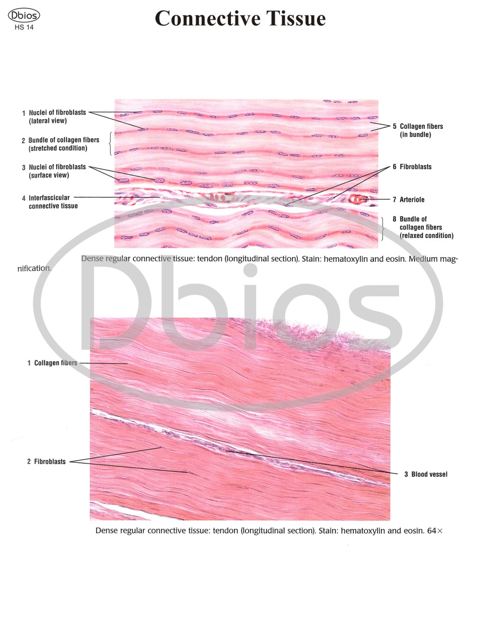
HO 03 Composite Illustration of Loose Connective Tissue with Its Predominant Cells and Fibers
HO 04 Endochondral ossification, Illustrating the Progressive Stages of Bone Formation(from Cartilage Model to Bone) and Including the Histology of A Section of Formed Bone
HO 05 Differentiation of A Pluripotential Hemopoietic Stem Into the Myeloid Stem Cell Line and Lymphoid Stem Cell Line During Hemopoiesis
HO 06 Microscopic Illustrations of the Three Types of Muscles: Skeletal, Cardiac, and Smooth
HO 07 The central nervous system is composed of the brain and spinal cord. A section of the brain and spinal cord is illustrated here with their protective connective tissue layers called meninges (dura mater, arachoid, and pia mater)
HO 08 The peripheral nervous system is composed of the cranial and spinal nerves. A cross – section of the spinal cord is illustrated here with the characteristic features of the motor neuron and a cross-section of a peripheral nerve. Also illustrated are types of neurons located in different ganglia and organs outside of the central nervous system
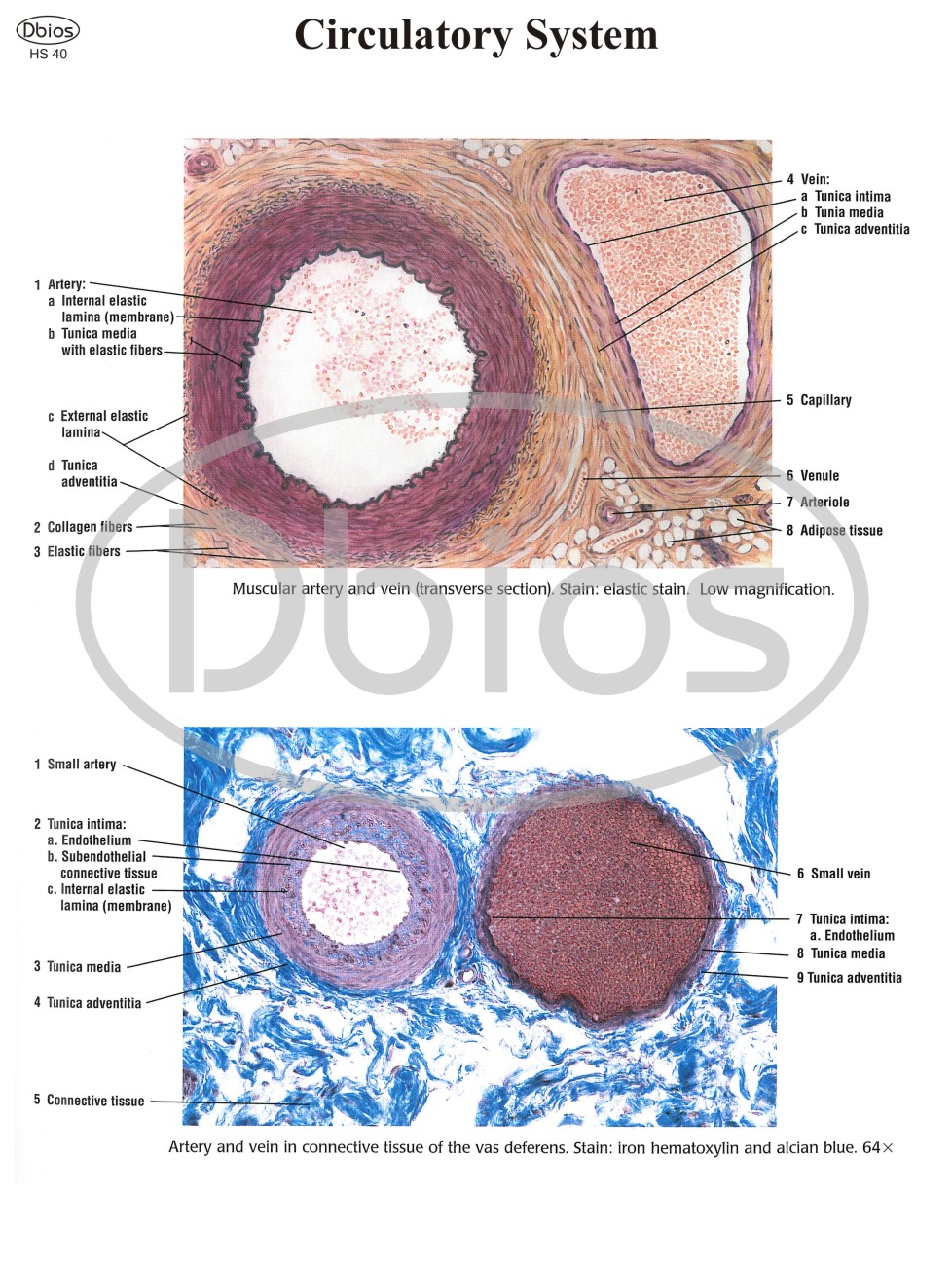
HO 09 Comparison (transverse sections) of a muscular artery, large vein, and the three types of capillaries
HO 10 Location and distribution of the lymphoid organs and lymphatic channels in the body. Internal contents of the lymph node and spleen are illustrated in greater detail
HO 11 Comparison between thin skin in the arm and thick skin in the palm, including contents of the connective tissue dermis
HS 01 Simple Squamous Epithelium: Peritoneal Mesothelium Surrounding Small Intestine Different Epithelial Types In the Kidney Cortex
HS 02 Simple Columnar Epithelium : Stomach Surface
HS 03 Simple Columnar Epithelium on Villi in Small Intestine : cells with Striated Borders(Microvilli) and Goblet Cells
HS 04 Pseudostratified Columnar Ciliated Epithelium: Respiratoy Passages (Trachea)
HS 05 Transitional Epithelium: Bladder (contracted)
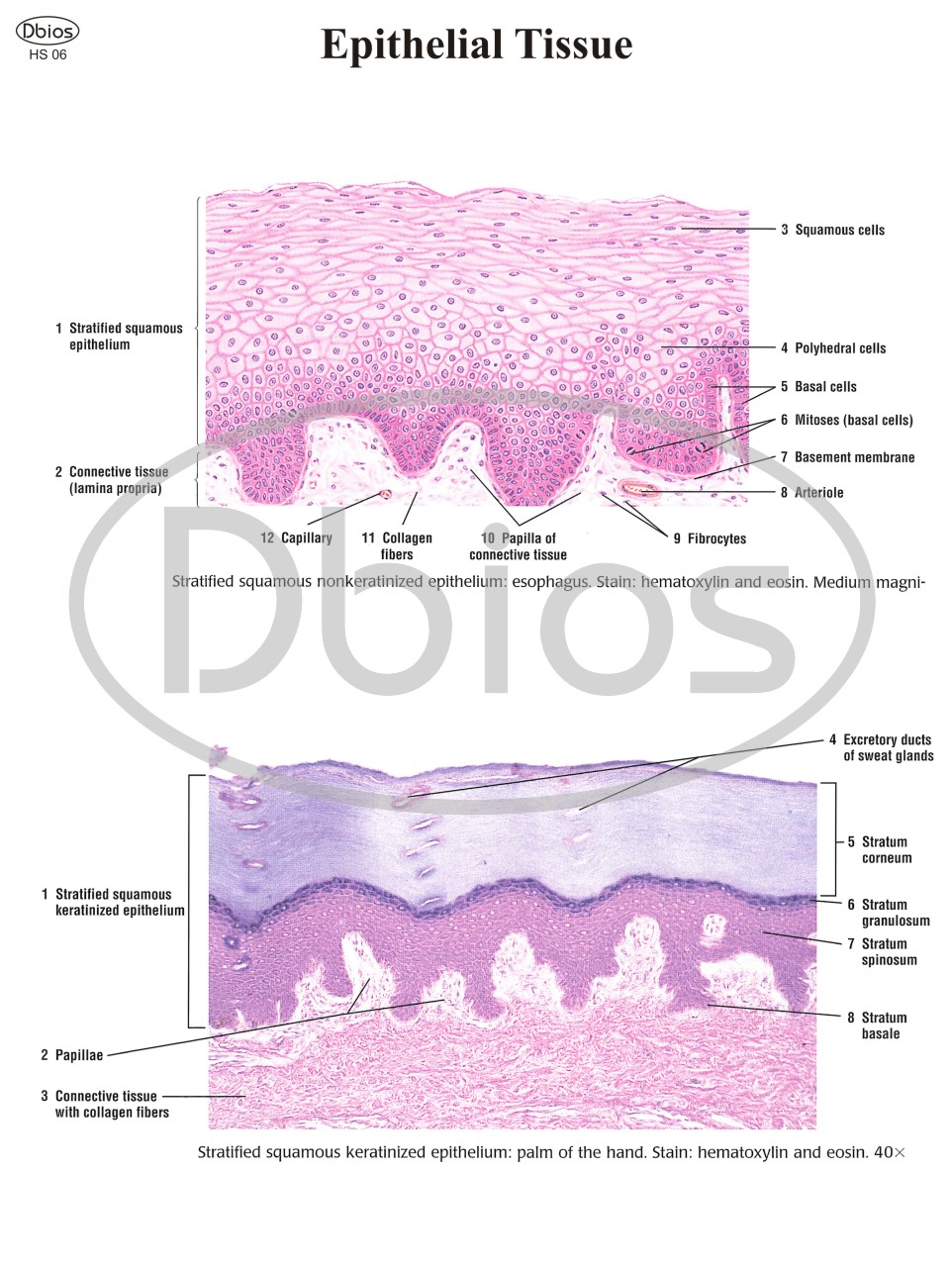
HS 06 Stratified Squamous Nonkeratinized Epithelium: Esophagus & Keratinized Epithelium: Palm of the Hand
HS 07 Stratified Cuboidal Epithelium: Excretory Duct In Salivary Gland Glandular Tissue
HS 08 Unbranched Simple Tubular Exocrine glands: intestinal glands Simple Branched Tubular Exocrine Glands: Gastric Glands
HS 09 Coiled Tubular Exocrine Glands: Sweat Glands & Compound Acinar (exocrine) Gland: Mammary Gland Connective Tissue
HS 10 Loose Connective Tissue
HS 11 Individual Cells of Connective Tissue
HS 12 Loose Connective Tissue & Dense Irregular and Loose Irregular Connective Tissue (Elastin Stain)
HS 13 Loose Irregular and Dense Irregular Connective Tissue Dense Irregular Connective Tissue and Adipose Tissue
HS 86 Ovary: Dog (panoramic View)
HS 87 Ovary: Ovarian Cortex and Primary and Primordial Follicles
HS 88 Uterine Tube:ampulla (panoramic View, Transverse Section) Uterine Tube: mucosal Folds (early Proliferative Phase)
HS 89 Uterus: Proliferative (follicular) Phase
HS 90 Uterus:secretory (luteal) Phase
HS 91 Uterus:menstrual Phase
Cervix, Vagina, Placenta, and Mammary Glands
HS 92 Vagina (longitudinal Section)
Glycogen In Human Vaginal Epithelium
HS 93 Vagina: Surface Epithelium Placenta At 5 (panoramic View)
HS 94 Inactive Mammary Gland Mammary Gland During Proliferation and Early Pregnancy Organs of Special Senses
HS 95 Eyelid (Sagittal Section)
HS 96 Lacrimal Gland & Cornea (Transverse Section)
HS 97 Whole Eye (Sagittal Section) & Retina,choroid, and Sclera (panoramic View)
HS 98 Layers of the Choroid and Retina (detail) & Eye: Layers of Retina and Choroid
HS 99 Inner Ear: Cochlear Duct (scala Media) & Inner Ear; Cochlear Duct and the Organ of Corti